The mitral valve separates the two chambers (atrium and ventricle) on the left side of the heart and keeps blood moving in the correct direction. If the valve does not close properly, blood may leak back from the ventricle into the atrium (known as regurgitation or insufficiency), which reduces the efficiency of the heart as a pump. If the valve is narrowed or rigid (known as stenosis)—for instance, due to calcium deposits or scarring—blood flow is obstructed and the heart must again work harder to pump blood through. Either condition, if severe, may eventually lead to heart failure, stroke, or heart attack.
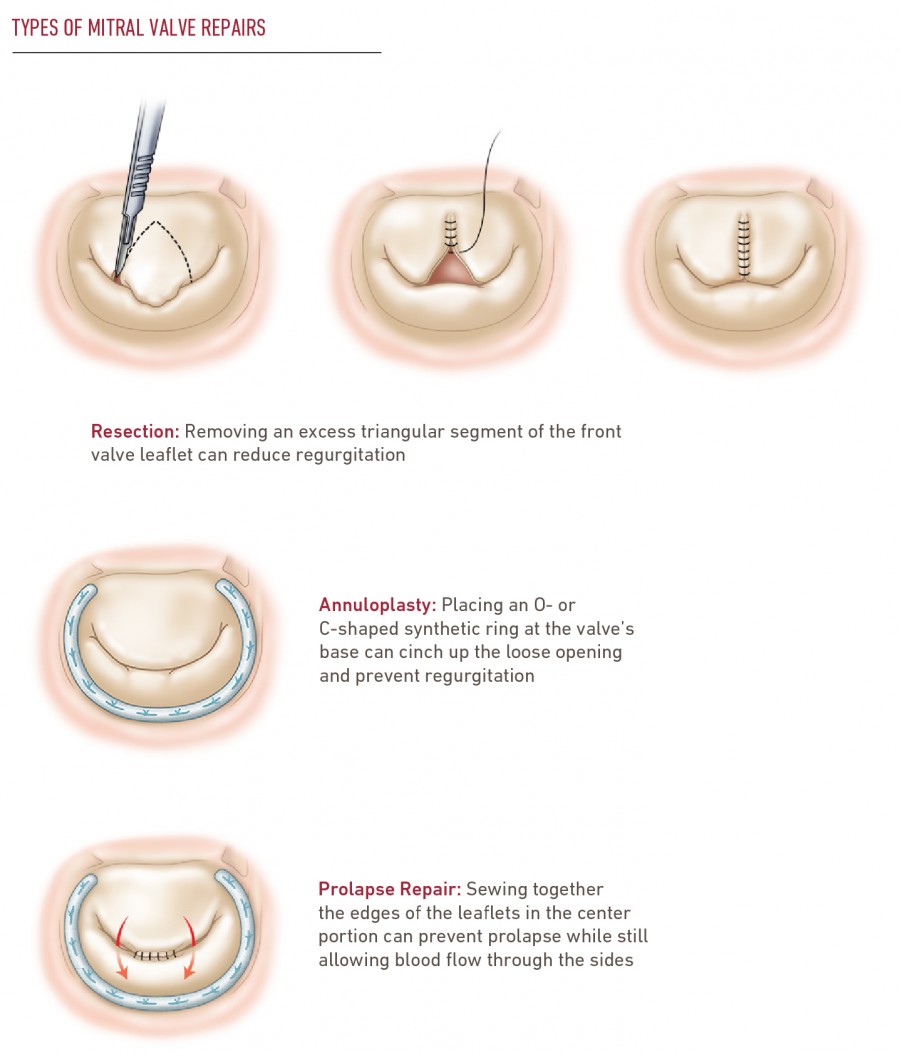
The goal of mitral valve surgery is to repair valve tissue or connecting structures so that the valve can open properly and seal tightly. For example, in some mitral valve repairs, your heart surgeon may separate fused leaflets, create better openings, or remove calcium or other obstructions. In others—for instance, to treat a weak and bulging (prolapsed) valve—your surgeon may trim valve leaflets and insert an annuloplasty device to reshape and strengthen the floppy mitral valve. If the valve is damaged beyond repair, it may be replaced with a new synthetic or donor-tissue valve. Many mitral valve procedures can be performed using minimally invasive techniques.
 Why Temple?
